Correctly interpreting the cause of chest pain is a crucial diagnostic skill that is essential for effective medical practice. Before the development of coronary angioplasty and stenting, the great majority of patients with stable angina (typical or atypical) were treated medically. Currently, patients with stable angina often undergo early invasive coronary angiography with an eye toward intervention for sufficiently stenotic lesions. The goal of this approach has been to eliminate angina rather than to reduce the risk of cardiovascular events.
This more aggressive approach has improved the lives of many patients with stable angina, to be sure. Yet, this strategy has not been without risks and challenges. First, a substantial percentage of patients (approximately 60%) who are referred for angiography have no hemodynamically significant coronary artery stenoses. Thus, many patients without coronary artery disease (CAD) are subjected to the low but real risk of complications from angiography. Second, defining the anatomy does not necessarily provide useful information about the hemodynamic importance of a lesion unless such definition is accompanied by some demonstration of impaired perfusion. For these reasons, a variety of noninvasive stress studies have been developed to aid in the identification of a flow-limiting lesion, each with reasonable sensitivity and specificity. Yet, among 52% of patients with abnormal results on functional testing, only approximately half were found to have obstructive CAD on subsequent angiography. These results suggest that alternate diagnostic strategies need to be considered to identify obstructive lesions of functional importance.
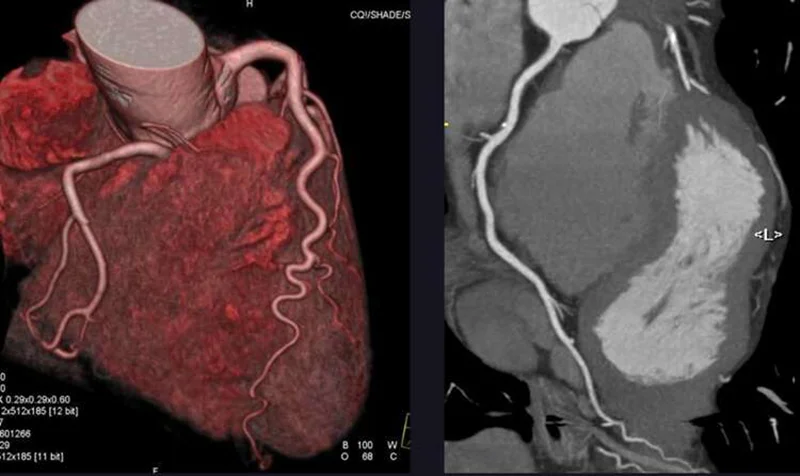
Over the past 20 years, coronary computed tomographic angiography (CCTA) has emerged as another noninvasive method for diagnosing obstructive CAD. Among patients with intermediate or high pretest probability of obstructive CAD, CCTA has been found to have a sensitivity, specificity, positive predictive value, and negative predictive value for identifying obstructive CAD of 0.92, 0.75, 0.84, and 0.87, respectively.5 On the basis of these data and the results of five randomized, controlled trials conducted over the past 10 years, CCTA has become the preferred imaging approach for the assessment of patients with stable chest pain, especially in those with an intermediate pretest probability of obstructive CAD.
The members of the DISCHARGE Trial Group now report in the Journalthe results of a pragmatic, randomized trial of CCTA as compared with angiography as an initial diagnostic imaging strategy in 3561 patients with stable chest pain and an intermediate pretest probability of obstructive CAD. The authors found no material difference between CCTA and angiography in the incidence of the primary composite outcome of cardiovascular death, nonfatal myocardial infarction, or nonfatal stroke during 3.5 years of follow-up. This result is probably a consequence of the lack of effect of revascularization on cardiovascular events among most patients with stable angina and the limited number of those with high-risk anatomy who would benefit from revascularization in the trial (13.9% in the CCTA group and 11.2% in the angiography group). In addition, CCTA was performed significantly earlier than angiography (3 days vs. 12 days after enrollment), which may have led to earlier revascularization and a better outcome in patients whose anatomy would benefit from it.
As with many trials that advance a field, this trial raises several additional questions of interest. Why was a pretest probability of obstructive CAD of 10 to 60% chosen as the cutoff for intermediate risk, and on what basis was the scoring system chosen to calculate that probability? Would a different range cutoff or a different scoring system have led to a different trial outcome? Was the inclusion of a large number of patients with nonanginal chest pain (>35%) justified, and would their exclusion have led to a different trial outcome? Do these two trial design features account for the fact that only approximately one quarter of the patients in each group had obstructive CAD, which suggests that the overall trial population had a low risk of obstructive CAD rather than an intermediate risk? The most recent guidelines of the American College of Cardiology–American Heart Association for the evaluation and diagnosis of chest pain recommend no testing and intensification of goal-directed medical therapy in low-risk patients.
From the functional-assessment perspective, would the incorporation of other noninvasive measures of impaired perfusion, such as CT-based estimates of fractional flow reserve, further improve the predictive accuracy of CCTA? Can CCTA-based quantitation of the plaque burden and lesion characterization improve predictive accuracy, especially given that many lesions leading to acute coronary events are not flow-limiting before plaque activation? No doubt, these and other questions will serve as the basis for future trials involving CCTA as its incorporation in the assessment of patients with stable angina continues to evolve.

دیدگاه خود را بنویسید