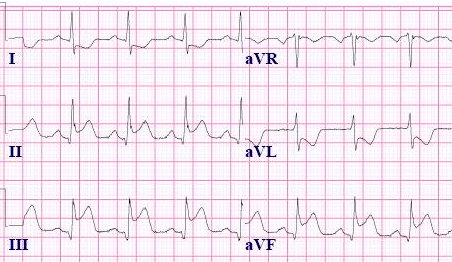
Fortunately, recognizing the inferior STEMI is a bit more straightforward. This MI involves ST segment elevation in the inferior leads II, III and aVF and only requires 1 mm in 2 contiguous leads. There is usually reciprocal depression in leads I and aVL, which helps to distinguish this from pericarditis. There is not a lot of variation in how an inferior MI looks in regards to shape or ST segments; however, some are more dramatic than others based on the amplitude of ST segment elevation. Also, during an inferior MI, the ST segment elevation is usually concave upwards.
Below are some examples to see what they look like.
Inferior STEMI Example #1
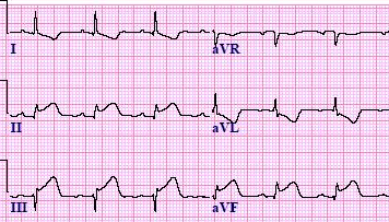
Inferior STEMI Example #2
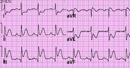
Inferior STEMI Example #3

Inferior STEMI Example #4
دیدگاه خود را بنویسید